Self-healing and Self-destruction – Part 1
Event data
- Datum
- 4. 4. 2017
- Host
- Mini-Med Studium
- Location
- Van-Swieten-Saal der Medizinischen Universität Wien
- Event-type
- Vortrag
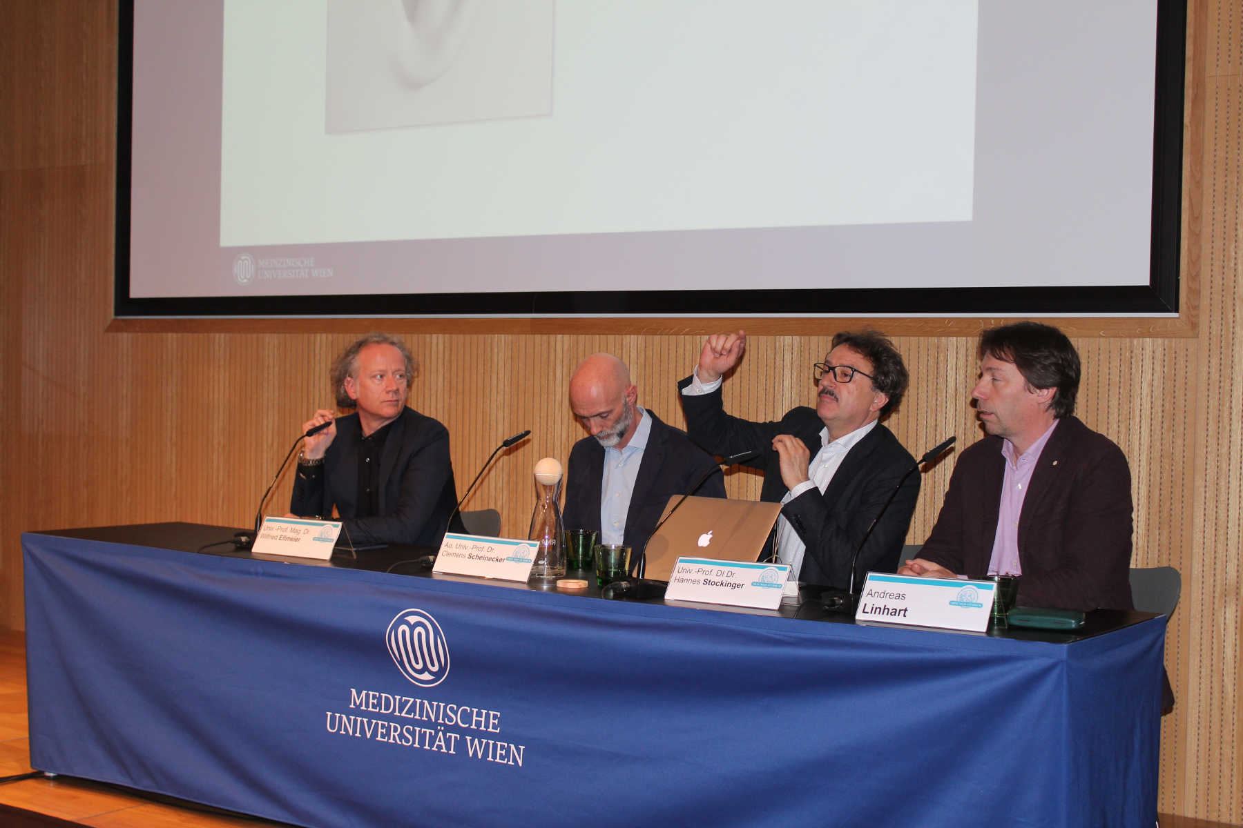
- Participants
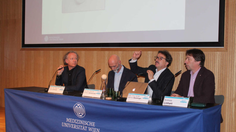
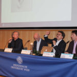
- Univ.-Prof. Mag. Dr. Wilfried Ellmeier, Institut für Immunologie, MedUni Wien
- Ao. Univ.-Prof. Dr. Clemens Scheinecker, Universitätsklinik für Innere Medizin III, MedUni Wien/AKH Wien
- Univ.-Prof. DI Dr. Hannes Stockinger, Institut für Hygiene und Angewandte Immunologie, MedUni Wien
On Tuesday, 4 April 2017, a further event in the Mini-Med-Reihe series was held in the Van-Swieten-Saal. ein weiterer Termin der Mini-Med-Reihe statt. Three professors, Wilfried Ellmeier, Clemens Scheinecker and Hannes Stockinger gave talks on the subject of the evening, “Fascinating Immune System: The Balance between Self-healing and Self-destruction”.
Definition of immune system and some figures
Prof. Ellmeier began his talk with an explanantion of the immune system, which consisits of immune cells. Blood is of particular importance for the immune system. Although blood is red due to the erythrocytes (the red blood cells responsible for the transport of oxygen, 85% of all cells in the human body), it also contains, among others, white blood cells, leukocytes. These protect us from pathogens as part of the immune system. As blood circulates throughout the body, these can also be found everywhere in the body. From the leukocytes, there are approx. 4,000-10,000/μl blood (in comparison: the corresponding figure for red blood cells is approx. 4-7 million/μl blood). If one projects these figures onto approx. 5-6 litres of blood, there are billions of cells. Lymphocytes, granulocytes and monocytes belong, among others, to the white blood cells in our circulation (so-called “blood immune cells”). Various organs also contain immune cells, including macrophages, dendritic cells and mastocytes.
In addition to blood (blood immune cells) there are, as mentioned, certain organs in which there are a multiplicity of immune cells. These include, for example, the lymph nodes and the spleen. Further, there are many immune cells in precisely those regions which have much contact with the environnment, such as the oral mucosa, the nose, the lungs and also the intestine. A further organ which is full of immune cells is the skin.
Tasks of the immune cells
The following belong to the immune cells:
- Neutrophils – it is possible for these to “eat up” pathogens (specialist term:”phagocytosis”).
- Macrophages – these are also important phagocytes.
- Dendritic cells – cells with appendages (“dendron“ from the Latin for tree)
- Lymphocytes – of which there are various types:
- B-lymphocytes – these produce antibodies produzieren Antikörper
- T-lymphocytes – to which the T-helper cells belong (help B-cells to produce antibodies) and T-killer cells (can kill other cells e.g. if the cell has a virus)
Formation of immune cells

And then what happens?
After they have been formed, the imune cells flow from the bone marrow and the thymus and wander to the various organs, e.g.:
- the lymphatic system
- blood vessels and the blood system
- the lymph nodes
- the spleen
- the intestine
It is possible for immune cells to recogise pathogens – T-lymphocytes and dendritic cells in particular (“antigen presenting cells”) play an important role here. It is possible for the dendritic cells to present fragments of the antigen on the surface (Author’s note: antigens are characteristics – normally protein, carbohydrate or fat and other complex molecules – on the surface of pathogens). Every T-lymphocyte which the antigen recognises then multiplies and can perform its functions.
Cell-cell communication
This is only possible via communication with other cells. Communication between cells is possible by means of the release of certain messenger substances (“cytokine”). These are soluble proteins. If there are cells present nearby which could recognise these proteins due to their surface receptors (“recipients”), communication is possible between the two. There are various kinds of information which can thus be exchanged:
- “Please wake up and work!”
- “Please divide yourselves!”
- “Please produce antibodies!”
- “Please stop, it is enough!”
Prof. Ellmeier concluded his talk by stressing that immunological research is one of the five research focus points at the Medical University in Vienna.
Prof. Hannes Stockinger spoke about specific defence mechanisms
The B- and T-lymphocytes belong to the innate and adaptive immune system.
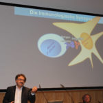
The immunological synapse
It is thought that the solution to these immune dysfunctions lies in the so-called “immunoligical synapse”. This is the “kissing zone” in which the antigen presenting cells (“dendritic cells”) present the T-lymphocytes to the antigen (surface characteristics). These require an “igniter” to react. The pathogen has to be processed by the dendritic cells in order to stimulate the T-lymphocytes to grow and to develop into T-killer cells.

The importance of the dendritic cells can be seen in the example of the skin. The cells are so dense that no pathogen can pentrate this barrier, nothing can pass through. In the case of injury and the consequent infiltration of pathogens, the dendritic cells react quickly in presenting the antigen of the pathogen and this leads to the immediate activation of the T-lymphocytes.
There has been quite some success in this area in the last few years. Different pathogens, but also cancer cells, “borrowed” these inhibitors and thereby attempted to inhibit the natural human immune cells. Since around 2013 the very successful cancer treatment of melanomas (“black skin cancer”), lung cancer as well as – currently on trial – colon cancer has followed the priniciple of inhibiting these inhibitors. As a result of the inhibition of the inhibitors, the T-lymphocytes begin to further develop into T-killer cells and start to destroy the cancer cells.
The receptors
To destroy the enemy, one has also to be able to recognise it. For this, all cells (e.g. macrophages, granulocytes and dendritic cells) have so-called recognition structures on the surface – these are termed “receptors”. The cells of the macrophages, the granulocytes and the dendritic cells have some “germline determining” (author’s note: already present in one’s genes before birth) receptors i.e. in our DNA there is a piece of a gene which is “transcribed” into a protein.
In the case of B and T-lymphocytes, this process is completely different and has not been understood for a long time, according to Prof. Stockinger. It is possible for these to produce an infinite number of diverse receptors. It is not clear how this functions. The B-lymphocytes are also able to emit their antigen receptors in soluble form, these then being called antibodies.
How is such an antibody released? This follows by a pathogen (to be more precise, the antigen on the pathogen).
There are, however, millions of pathogens – how is it possible for the B and T-lymphocytes to produce so many different antigen receptors? A person has “only” 22,000 genes and as such a limited genome.
Who revealed this secret of how it is possible for the B and T-lymphocytes to achieve a much greater antigen receptor variability – this and much more will be examined in the second part of the human immune system.